Anatomy Series

Ulnar Collateral Ligament Sprain

Elbow Stability and Movement Coordination Impairments

Screen Shot 2018-11-03 at 2.25.21 PM.png
  • Ulnar collateral ligament (UCL) pathology often occurs in individuals who perform repeated/forceful overhead movements. If your patient is an overhead athlete complaining of medial elbow pain, check the UCL! 

  • If you do not know the common clinical findings no problem! Click here


Anatomy

Picture1.png

Image via Complete Anatomy 2018 by 3D4 Medical


Key Finding

Palpation of the UCL can help to assess if the UCL is indeed a pathological source tissue. If radicular symptoms are present upon palpation, especially in in the 4th and 5th digits, there could be ulnar nerve involvement as well! (Click image to watch 1-2 minute video)

Movement Fault

In overhead athletes, decreased shoulder external rotation range of motion can result in increased valgus stress directed at the UCL! (Click image to watch 1-2 minute video)

Clinical Pearl: In overhead athletes it is important to assess the entire kinetic chain in order to determine if faulty force transmission is present. For example, research shows that decreased hip internal rotation range of motion can also result in excessive force transmission through the shoulder and the elbow!

Special Test

The moving valgus stress test is excellent for ruling out UCL pathology with a sensitivity of 100! Take a look and see how its done! (Click image to watch 1-2 minute video)

Treatment/Therapeutic Exercise


Research suggests that in overhead athletes, the posterior aspect of the glenohumeral joint capsule can often contracture due to high rates of repetitive movement. This change in tissue can result in limited shoulder range of motion and subpar shoulder arthorokinematics potentially leading to increased strain on structures like the UCL! The patient can also be taught how to perform this exercise at home! (Click image to watch 1-2 minute video)

*Acutely, addressing pain and inflammation around the UCL via techniques such as soft tissue mobilization and activity reduction may prove to be beneficial.

Post Traumatic Elbow Stiffness

Elbow Pain with Mobility Deficits

Screen Shot 2018-11-03 at 2.56.40 PM.png
  • Often, after traumatic injury (e.g. fracture) or immobilization, the joints of the elbow along with the joint capsule can become stiff resulting in pain and decreased range of motion! 

  • If you do not know the common clinical findings no problem! Click here


Anatomy

Picture1.png

Image via Complete Anatomy 2018 by 3D4 Medical


Key Finding

If a patient presents to clinic with elbow pain and decreased range of motion, check for history of trauma/immobilization! (Click image to watch 1-2 minute video)

Mobility Assessment

Post immobilization, elbow extension tends to be the most limited. Although the ulnohumeral joint is responsible for the majority of elbow extension, it is imperative to assess the radiohumeral and proximal radioulnar joint as well! (Click image to watch 1-2 minute video)

Treatment

Ulnohumeral distraction mobilization can help improve both flexion and extension range of motion! (Click image to watch 1-2 minute video)

Therapeutic Exercise


Once you have improved joint mobility it is important to follow up with movement in the new found range of motion! (Click image to watch 1-2 minute video)

Clinical Pattern Recognition - Shoulder Pain

From anatomy to discovering the patient!

Shoulder pain.jpg
  • Primary shoulder dislocation peaks in the second and sixth decade

  • 98% of cases, shoulder displaces anteriorly

  • 70% of people who have dislocated can expect to dislocate again within 2 years of the initial injury

  • Sub-acromial pain syndrome accounts for 44% to 60% of all conditions that cause shoulder pain and is the most frequent cause of visits to a physician’s office

  • See more prevalence information in the Clinical Pattern Recognition: Orthopaedics app here

Meet the 5 common shoulder pain patients from the Shoulder Pain and Mobility Deficit Clinical Practice Guidelines and more!


Clinical Pattern Recognition

Click on the pain pattern to learn about the patients and develop your clinical patterns!

1. Adhesive Capsulitis or Frozen Shoulder Shoulder pain and mobility deficits (1-minute video)

2. Labral tears, SLAP lesions, Bankart lesions, Anterior or Multidirectional Instability - Shoulder pain with movement coordination impairments (1-minute video)

3. Sub-Acromial Pain Syndrome (previously Shoulder Impingement) - Shoulder pain and muscle power deficits (1-minute video)

4. Acromioclavicular Joint Sprain Shoulder pain and movement coordination impairments (1-minute video)

5. Thoracic Outlet Syndrome (TOS) Shoulder pain with radiating pain (1-minute video)

Adhesive Capsulitis (Frozen Shoulder)

Shoulder Pain with Mobility Deficits

  • The exact cause of adhesive capsulitis is not completely understood; however, it has been divided into primary and secondary categories; primary being idiopathic and secondary being related to systemic, extrinsic or intrinsic pathology. If your patient has a history of diabetes mellitus, thyroid dysfunction, or a history of contralateral frozen shoulder along with symptoms similar to 1 of the 4 clinical stages they may be suffering from adhesive capsulitis!

  • If you do not know the common clinical findings no problem! Click here


Sub-Acromial Pain Syndrome (Shoulder Impingement)

Shoulder Pain with Muscle Power Deficits

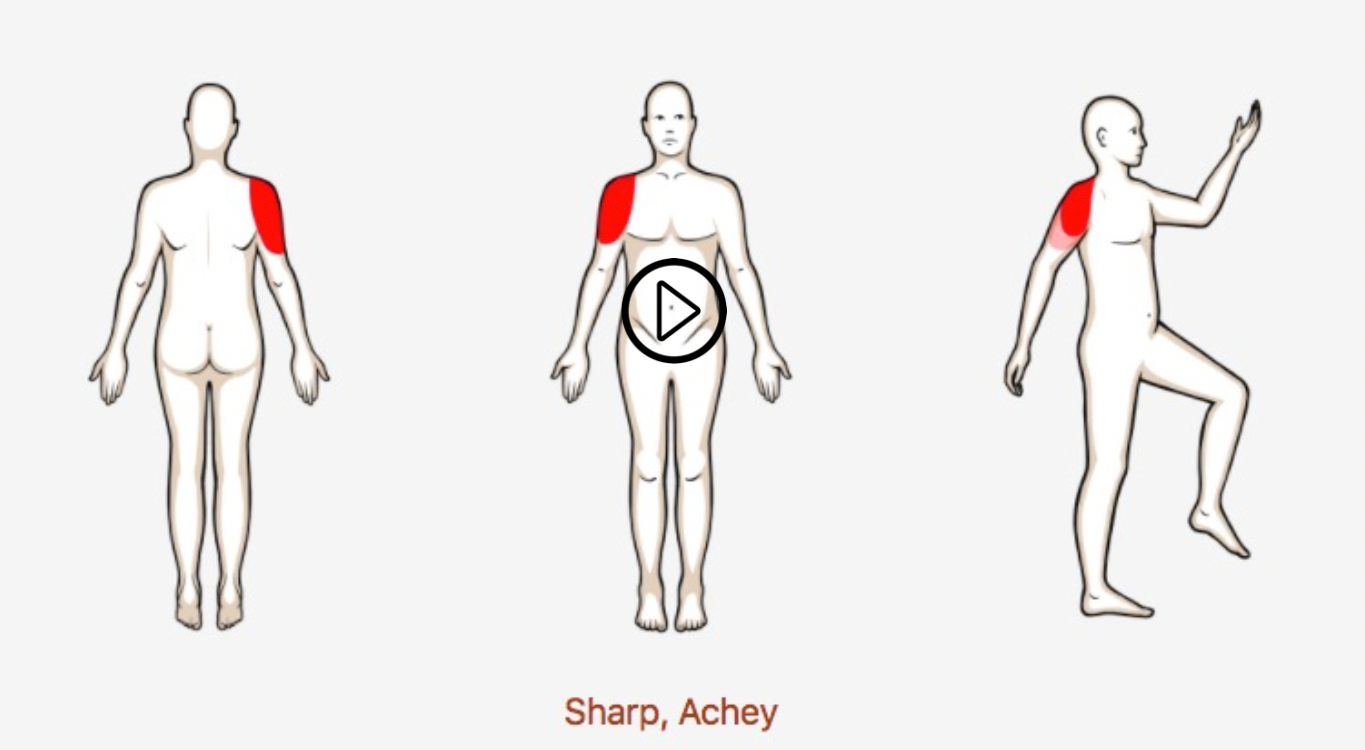
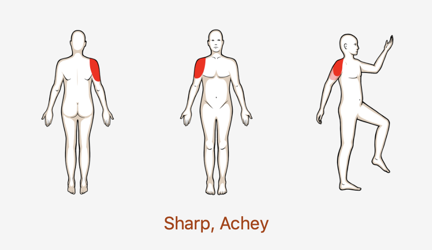
  • The Sub-acromial pain syndrome is perhaps the most common cause of shoulder pain in patients and a frequent cause of primary care physician visits. Patients will often present with sharp shoulder pain exacerbated in mid ranges of shoulder movement and repetitive activities. Take a look at some ways to assess and treat this common pathology!

  • If you do not know the common clinical findings no problem! Click here