Anatomy Series

Thoracic Kyphosis/Spondylosis

Mid Back Pain with Mobility Deficits

Screen Shot 2019-08-20 at 9.14.26 PM.png
  • Patients with excessive thoracic spine kyphosis often report diffuse achy pain in the mid thoracic spine; especially with upper trunk extension or extension-rotation movements. It is important to note that mobility deficits in the thoracic region can result in pain in surrounding structures such as the neck, shoulder, or lower back! For more clinical findings click here!


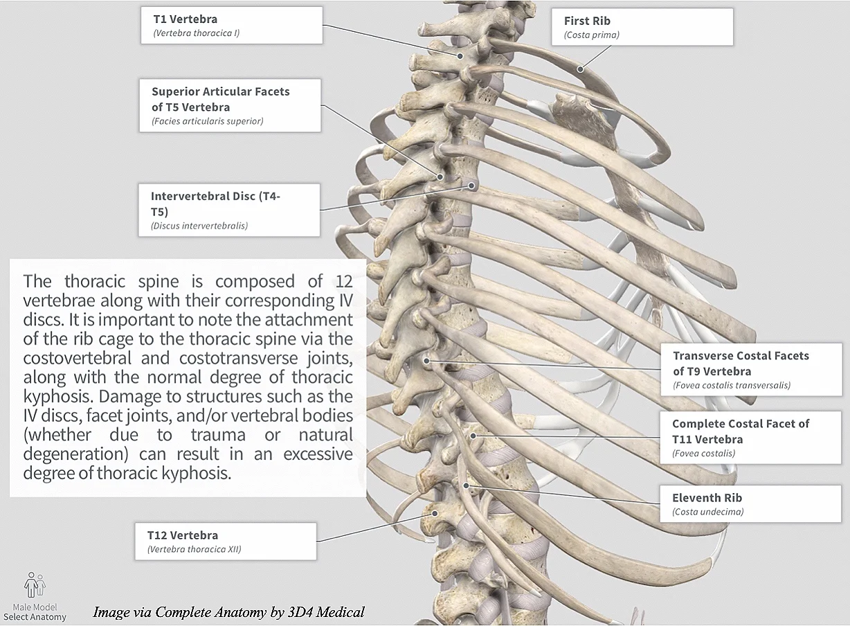
Anatomy

Image via Complete Anatomy by 3D4 Medical

Image via Complete Anatomy by 3D4 Medical


Movement Fault

  • Although excessive thoracic spine kyphosis can typically be identified in standing and/or seated postural assessments, the quadruped position is an excellent functional alternative to analyzing the thoracic spine and how it’s movement interacts with the rest of the body! (Click image to watch 1-2 minute video)

Key Finding

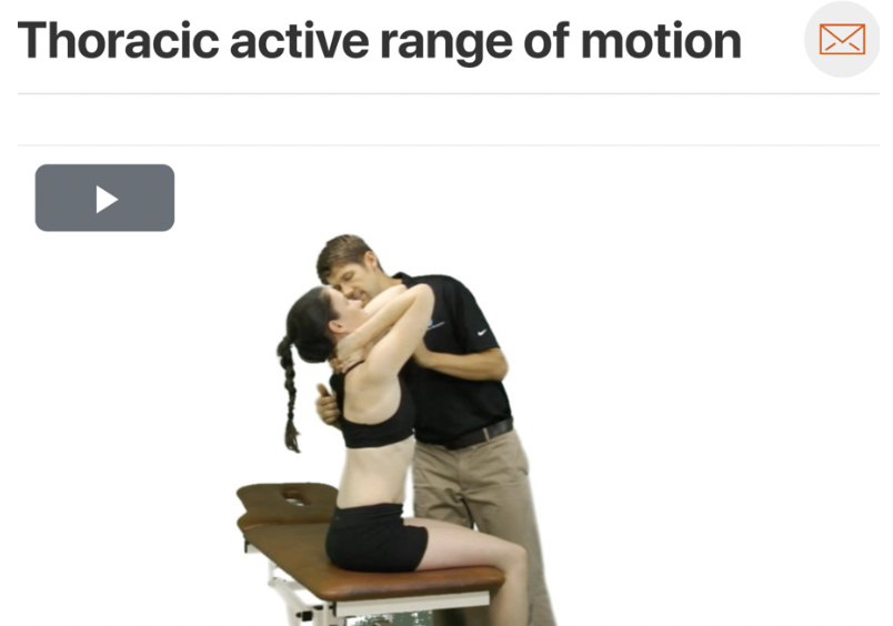
As mentioned before, patients with mobility deficits of the thoracic spine often experience exacerbation of symptoms with extension and/or extension rotation movements. Be sure to assess the patient’s active range of motion to see which direction they are most limited in! (Click image to watch 1-2 minute video)

Treatment

  • If the patient is deemed appropriate for manipulative therapy, a high velocity low amplitude thrust (HVLAT) can help provide immediate symptom improvement. (Click image to watch 1-2 minute video)

Therapeutic Exercise

  • After manual therapy is provided, it is imperative to follow up with therapeutic exercise in order to promote the new movement pattern. Although multiple exercises can help improve thoracic spine extension, it is important to give patients something they can do regularly. Thoracic spine extension over a chair is an exercise that can be performed anywhere! (Click image to watch 1-2 minute video)

  • As always, address the individual as a whole, determine which are the primary impairments and begin treatment there!

Sacroiliac Joint Sprain

Pelvic Girdle Mobility Deficits

Screen Shot 2019-08-20 at 9.14.26 PM.png
  • Various pathoanatomic and/or kinesiopathologic reasons exist for unilateral lower back/buttock pain; however, if a patient presents to clinic after sustaining a fall onto the buttock, abrupt misstep on a straight leg, or becoming pregnant, the sacroiliac joint (SIJ) may be the culprit! For more clinical findings click here!


Anatomy

Image via Complete Anatomy by 3D4 Medical

Image via Complete Anatomy by 3D4 Medical


Key Finding

  • Assessment of pelvic girdle alignment can help the clinician discover obliquities, which may be contributing to patient symptomology. (Click image to watch 1-2 minute video)

Special Test

The posterior pelvic pain provocation test (P4) is part of a cluster of examinations that can help the clinician rule in or rule out SIJ pathology! (Click image to watch 1-2 minute video)

Treatment

  • If hypomobility of the SIJ is a contributing factor to patient symptomology, regional manipulation can help improve mobility and potentially stimulate under-active musculature while gaiting pain! (Click image to watch 1-2 minute video)

Therapeutic Exercise

  • After optimal pelvic alignment has been attained, it is important to promote continued stability via force closure utilizing muscles such has the hip abductors and adductors! (Click image to watch 1-2 minute video)

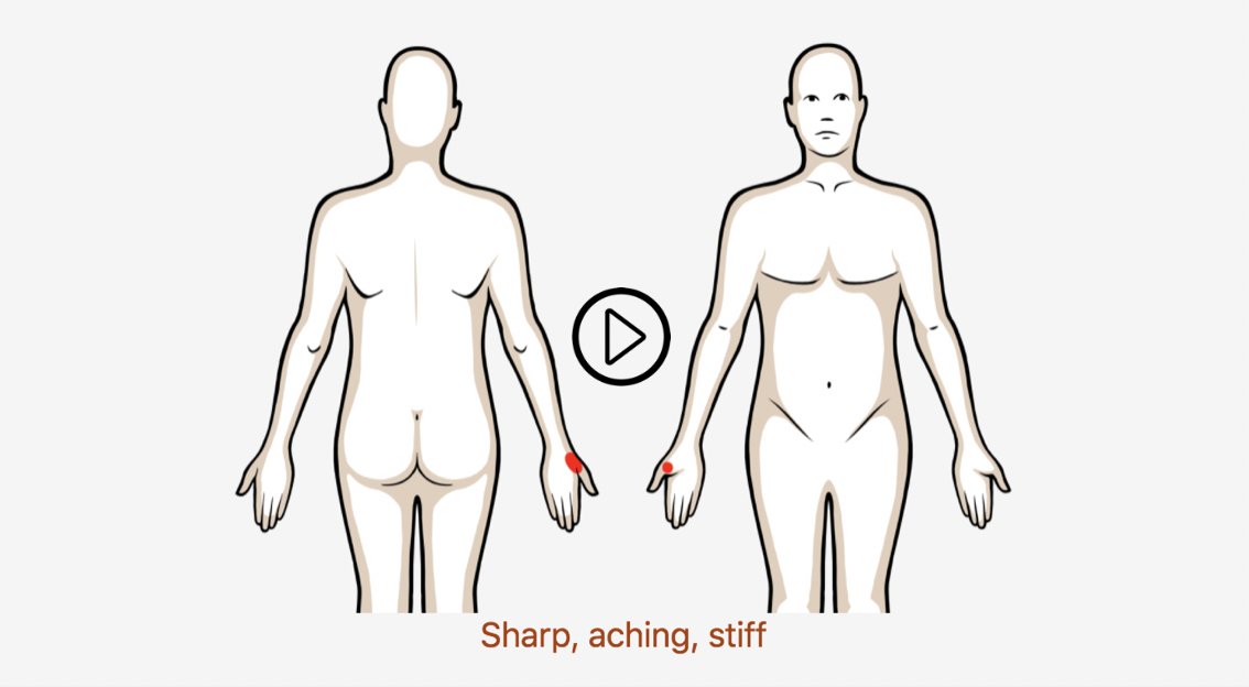
Clinical Pattern Recognition - Wrist/Hand Pain

From anatomy to discovering the patient!

Screen%252BShot%252B2019-08-19%252Bat%252B3.36.36%252BPM.jpg
  • Carpal tunnel syndrome is the most common upper extremity compressive neuropathy

    o 1 to 5% of general population

  • There were 11,332 cases of DeQuervain’s tenosynovitis in the population at risk of 12,117,749 person-years

  • Women had a significantly higher rate of DeQuervain’s tenosynovitis at 2.8 cases per 1000 person-years, compared to men at 0.6 per 1000 person-years

  • Triangular fibrocartilage complex (TFCC) was torn in 46 of 118 patients with distal radial

    fracture

    o 35% of intra-articular fractures

    o 53% of extra-articular fractures

  • In 59 hands with carpal tunnel syndrome, 34% also had ulnar tunnel (Guyon’s canal) neuropathy

See more prevalence information in the Clinical Pattern Recognition: Orthopaedics app here


Clinical Pattern Recognition

Click on the pain pattern to learn about the patients and develop your clinical patterns!

  1. Carpal Tunnel SyndromeHand Sensory Deficits (1-minute video)

2. DeQuervain’s Syndrome-Thumb Pain with Muscle Power Deficits (1-minute video)

3. Sprain of Carpal Ligaments–Wrist and Hand Pain with Movement Coordination Impairments (1-minute video)

4. Sprain of Ulnar Collateral Ligament of the ThumbThumb Pain with Movement Coordination Impairments (1-minute video)

5. Sprain or Tear of Triangular Fibrocartilage of the Distal Radius and UlnaWrist and Hand Pain with Movement Coordination Impairments (1-minute video)

6. Thumb Osteoarthritis- Thumb Pain with Mobility Deficits (1-minute video)

7. Ulnar Tunnel Syndrome or Entrapment Around Pisohamate Ligament- Hand Sensory Deficits (1-minute video)

8.  Wrist OsteoarthritisWrist Pain with Mobility Deficits (1-minute video)

Prevalence

Screen+Shot+2018-10-24+at+8.01.30+PM.jpg
  • Primary shoulder dislocate peaks in the second and sixth decade

  • 98% of cases, shoulder displaces anteriorly

  • 2% of cases, shoulder displaces posteriorly

  • 95% of first-time shoulder dislocations result from either a forceful collision, falling on an outstretched arm or a sudden wrenching movement

  • 5% of dislocations have an atraumatic origin (minor incidents such as raising the arm or moving during sleep

  • 70% of people who have dislocated can expect to dislocate again within 2 years of the initial injury

  • Younger and older subjects have a comparable incidence of primary shoulder dislocation

  • Incidence of recurrent dislocation is much more frequent in adolescent population

  • Dislocation reported to recur in:

    • 66% to 100% of people aged 20 years or under

    • 13% to 63% of people aged between 20 and 40 years old

    • 0% to 16% of people aged 40 years or older