Anatomy Series

Whiplash

Neck Pain with Movement Coordination Impairments

Screen Shot 2019-08-19 at 4.47.52 PM.png
  •  If a patient presents to clinic after a traumatic event such as a motor vehicle accident with neck pain and potentially back, shoulder, or arm pain, it is likely that they suffer from whiplash syndrome! For more clinical findings click here!


Anatomy

Screen Shot 2019-08-19 at 4.49.29 PM.png

Special Test

  • Research shows that motor control of the deep neck flexors is impaired in individuals with whiplash. The craniocervical flexion test is an excellent and reliable exam for evaluating deep neck flexor function Don’t forget to look for compensations! (Click image to watch 1-2 minute video)

  • Clinical Side Note: If cervical spine ligamentous instability or vertebral artery insufficiency cannot be ruled out during subjective examination, it is imperative to assess for these conditions prior to further cervical spine examination! (Click here to see how this is done) 

Treatment/Therapeutic Exercise

  •  If deep neck flexor endurance proves to be insufficient then the craniocervical flexion test or deep neck flexor endurance test can become a treatment option. Research also demonstrates that individuals with whiplash have impaired proprioception with respect to cervical movement. Use of pillows to unload sensitive structures while performing gentle range of motion exercises can help to improve this proprioceptive deficit! (Click image to watch 1-2 minute video)

Education

Education is perhaps the most important part of the rehabilitation process for individuals with whiplash. Considering that research demonstrates a significant likelihood of chronic pain occurrence in individuals with whiplash secondary to a motor vehicle accident, it is imperative that the patient is educated properly in order to intervene in this cycle. (Click image to watch 1-2 minute video)

Neck Sprain/Strain

Neck Pain with Movement Coordination Impairments

Screen Shot 2019-08-19 at 4.47.52 PM.png
  • Neck sprain/strains can occur due to a variety of factors including posture, ergonomic set up, excessive loading, and trauma. If your patient complains of tightness or spasm in the neck region along with difficulty maintaining static postures, a neck sprain/strain could be the culprit! For more clinical findings click here!


Anatomy

Screen+Shot+2019-08-19+at+5.03.22+PM.jpg

Key Finding

  • The upper trapezius can often times be a tissue source in patients with a neck sprain/strain. Lengthening, contracting, and palpating the suspected muscle can help the clinician better rule in the likelihood of strain! Take a look at how to assess upper trapezius length. (Click image to watch 1-2 minute video)

  •  Clinical Side Note 1: Considering that the upper trapezius and levator scapulae share similar locations, assessment of the levator scapulae may prove to be beneficial as well. 

Treatment

 Often times the upper trapezius (or levator scapulae for that matter) becomes irritated, not necessarily due to overactivity in a shortened position, but due to overactivity in a lengthened position secondary to variables such as poor posture. This forced activation in a lengthened position can eventually result in irritation of the tissue over time. The purpose of the post isometric relaxation technique is not to stretch the muscle, but to help relax it. Take a look at how its done! (Click image to watch 1-2 minute video)

Therapeutic Exercise

  • As mentioned previously, the middle trapezius is one of the muscles that is often under-active in individuals with a neck sprain/strain. Strengthening of the scapulothoracic musculature can assist with supporting the strained tissues and further reduce the amount of strain over time! (Click image to watch 1-2 minute video)
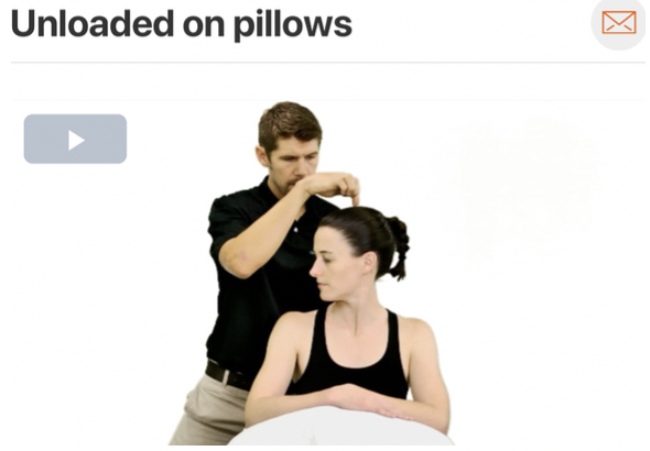
  • Clinical Side Note 2: Postural impairments tend to be present in these individuals. Do not forget to address posture and ergonomics. Make a functional change in something they are doing daily!

  • Clinical Side Note 3: Research also supports the use of thoracic spine manipulation (if deemed appropriate) in individuals with neck pain! If thoracic spine hypomobility is considered to be an associated impairment, consider adding this to the treatment plan!  

Cervicogenic Headache

Neck Pain with Headache

Screen Shot 2019-08-19 at 5.25.32 PM.png
  • When a patient presents with headache symptomology it is important for the clinician to be able to differentiate between the various types of headaches and treat accordingly. Cervicogenic headaches tend to be more commonly seen in clinic and typically present with unilateral sharp pain originating in the suboccipital region, which can travel up the cranium and toward the eye or down toward the neck! 

    For more clinical findings click here!


Anatomy

Image via Complete Anatomy by 3D4 Medical

Image via Complete Anatomy by 3D4 Medical


Key Finding

  • The cervical flexion rotation test is an excellent assessment to assist with headache differential diagnosis and identification of C1-C2 dysfunction. If impairment is present, it can also become a treatment! (Click image to watch 1-2 minute video)
Treatment

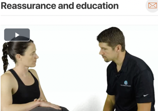
 If upper cervical hypomobility is present and the patient is deemed appropriate for thrust manipulation a 0-C1 distraction manipulation can help to improve range of motion and reduce pain. Take a look at how its done! (Click image to watch 1-2 minute video)

Therapeutic Exercise

  •  Cervicogenic headaches are often a result of posture and ergonomics with cervical extension and extension rotation movement faults being the most common. With these movement faults the suboccipital muscles can become short resulting in irritation. (Click image to watch 1-2 minute video)

  •  Clinical Side Note: If postural faults or poor ergonomics are present, do not forget to address them with your patient! 

Thumb Osteoarthritis

Thumb Pain with Mobility Deficits

Screen Shot 2019-08-19 at 5.25.32 PM.png
  • Do you or a family member have pain in your thumb? Do they complain of stiffness in the morning? Osteoarthritis of the thumb is commonly caused by injury, overuse injuries, or from a history of immobilization.  Look below for some things to consider!

    For more clinical findings click here!


Anatomy

Image via Complete Anatomy by 3D4 Medical

Image via Complete Anatomy by 3D4 Medical


Special Tests

  • A good clinical test you can use to check the integrity of the 1stCMC joint is the grind test. A positive test indicates osteoarthritis of the thumb. (Click image to watch 1-2 minute video

Treatment

 This technique can be used for both pain modulation and to help increase flexion range of motion in the thumb! (Click image to watch 1-2 minute video

Therapeutic Exercise

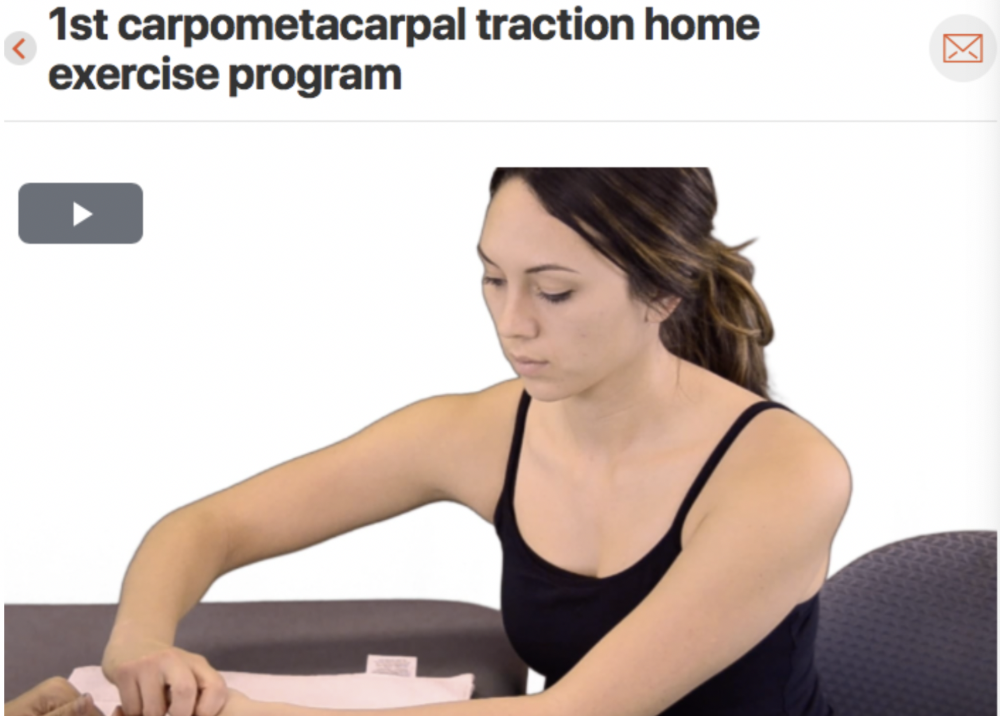
  • This exercise can be used to help mobilize the thumb at home and help decrease pain.  (Click image to watch 1-2 minute video

Carpal Tunnel

Wrist Pain with Radiating Pain

Screen Shot 2019-08-19 at 5.25.32 PM.png
  • Do you or a family member have numbness and tingling in your hand? Does it get worse when you are typing at work or while you are driving? If that is true, there is a chance that they are suffering from carpal tunnel.  Carpal tunnel is caused from increased pressure put on the median nerve as it runs through the carpal tunnel. If a patient presents with carpal tunnel like symptoms it is important to look further up the chain and make sure that there are not proximal entrapment sites causing your patients problems. Look below for some things to consider!

    For more clinical findings click here!


Anatomy

Image via Complete Anatomy by 3D4 Medical

Image via Complete Anatomy by 3D4 Medical


Special Tests

  • Patients with carpal tunnel often report a lessening of symptoms when they flick their wrist back and forth. They commonly will complain of symptoms when sleeping at night, have numbness and tingling in their first two fingers, and have difficulty picking up small objects. (Click image to watch 1-2 minute video

Treatment

This technique can be to decrease entrapment of the median nerve in the carpal tunnel. Make sure to follow this treatment with neural flossing!  (Click image to watch 1-2 minute video

Therapeutic Exercise

  • Nerve sliders can be used to decrease neural tension. Make sure to progress your patient to neural tensioners when appropriate. This exercise can be used to decrease sensitivity and improve mobility of the median nerve. (Click image to watch 1-2 minute video