Anatomy Series

Disc Herniation

Lower Back Pain with Radiating Pain

Screen Shot 2018-10-23 at 7.54.34 PM.png
  • Not to be confused with lower back pain with related leg pain; patients with a disc herniation will be more likely to have complaints of narrow shooting/burning type pain sometimes associated with numbness. It is important for the Physical Therapist to be able to make the distinction between the two.

  • If you do not know the common clinical findings no problem! Click here


Anatomy

Picture1.png

Image via Complete Anatomy 2018 by 3D4 Medical


Common Movement Fault

Similar to disc disorders due to displacement, typical mechanism of injury associated with discogenic herniation is bending forward to lift (often times heavy) objects. If the patient moves too readily through the lumbar spine and not enough through the hip (flexion movement fault), they are at a higher risk for back injury! (Click image to watch 1-2 minute video)

Special Tests


The straight leg raise is an excellent for ruling out disc herniation due to it’s high sensitivity (0.97)! (Click image to watch 1-2 minute video)

Treatment

It is important to note that in the early stages of disc herniation rehabilitation, the centralization procedures used for treating lower back pain with related leg pain may actually cause symptom exacerbation. Patient’s who present to clinic with an acute injury or high irritability may benefit from finding positions of comfort, maintaining neutral spine alignment, and slow restoration of normal lumbar spine lordosis. (Click the image to watch 1-2 minute video)

Therapeutic Exercise

The quadruped rock back is an excellent exercise for this patient population. It puts the lumbar spine in a gravity eliminated position and teaches the patient how to move through their hips while maintaining a neutral lumbar spine! (Click image to watch 1-2 minute video)

Discogenic Lower Back Pain

Lower Back Pain with Related Leg Pain

Screen Shot 2018-10-23 at 9.28.32 PM.png
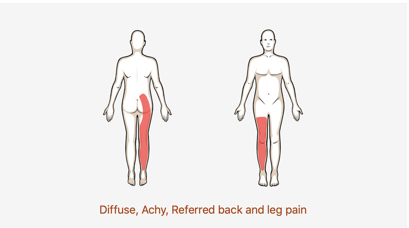
  • Is your patient experiencing diffuse or difficult to pin point pain that can travel down their leg? Take a look at some common clinical findings for discogenic lower back pain to see if signs and symptoms match up!

  • If you do not know the common clinical findings no problem! Click here


Anatomy

Picture1.png

Image via Complete Anatomy 2018 by 3D4 Medical


Common Movement Fault

Typical mechanism of injury associated with discogenic lower back pain is bending forward to lift (often times heavy) objects. If the patient moves too readily through the lumbar spine and not enough through the hip (flexion movement fault), they are at a higher risk for back injury! (Click image to watch 1-2 minute video)

Special Tests


The above videos demonstrate how you can quickly screen for discogenic lower back pain via centralization phenomenon! (Click images (Left/Right) to watch 1-2 minute video)


Treatment/Therapeutic Exercise

Repeated extension in standing can be used as both a treatment and a therapeutic exercise that the patient can perform on their own. If patient is unable to perform in standing try prone on elbows! Lastly, do not forget to address the flexion movement fault not just in standing but sitting as well! (Click the images (Left/Right) to watch 1-2 minute video)

Clinical Pattern Recognition- Ankle & Foot Pain

From anatomy to discovering the patient!

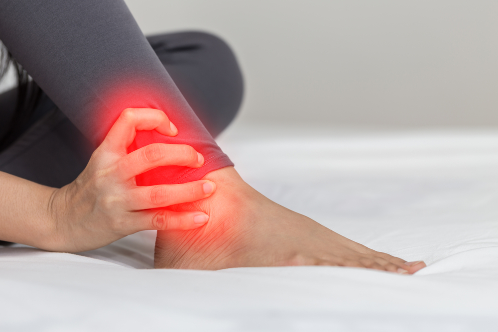
Ankle foot pain.jpg
  • Ankle joint accounts for 10% to 34% of all sport-related injuries, with lateral ankle sprain comprising 77% to 83% of these injuries
  • The overall incidence of lateral ankle sprain may be underestimated because approximately 50% do not seek medical attention after injury
  • 100% of 117 therapists who responded noted plantar fasciitis was the most common foot condition seen in the clinic
  • In the athletic population, plantar fasciitis is a common injury reported by both high school, competitive and recreational distance runners

See more prevalence information in the Clinical Pattern Recognition: Orthopaedics app here

Meet the 8 common ankle/foot pain patients from the Achilles Tendonitis Clinical Practice Guidelines, Ankle Stability and Movement Coordination Clinical Practice Guidelines, Heel Pain Clinical Practice Guidelines, and MORE!


Clinical Pattern Recognition

Click on the pain pattern to learn about the patients and develop your clinical patterns!

2. Ankle arthrosis- Ankle pain and mobility deficits (Watch 1 min video)

2. Achilles tendinosis- Ankle stiffness and muscle power deficits (Watch 1 min video | Step-by-step Guide)

3. Tibialis anterior tendonitis/Medial tibial stress syndrome- Leg pain and muscle power deficits (Watch 1 minute video)

4.  Ankle sprain- Ankle stability and movement coordination impairments (Watch 1 min video | Step-by-Step Guide)

5. Hallux rigidus- Great toe pain and mobility deficits (Watch 1 min video)

6. Posterior tibialis tendinitis/tendinosis- Ankle pain and muscle power deficits (Watch 1 min video | Step-by-Step Guide)

7. Plantar fasciitis - Heel pain (Watch 1 min video | Step-by-Step Guide)

8. Tarsal tunnel syndrome- Ankle and foot radiating pain (Watch 1 min video)

Achilles Tendinopathy

Muscle Power Deficits

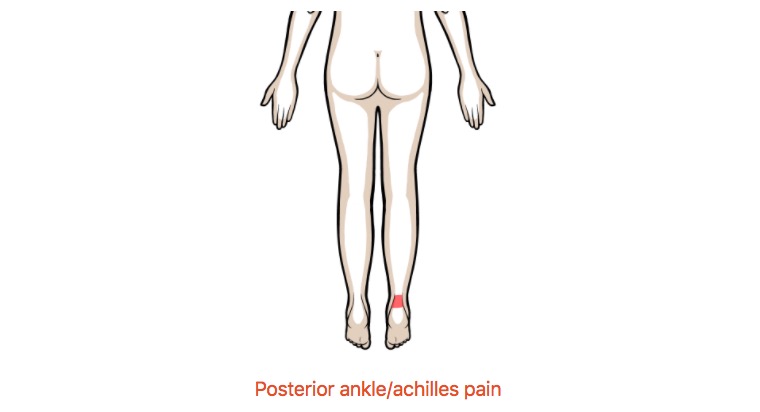
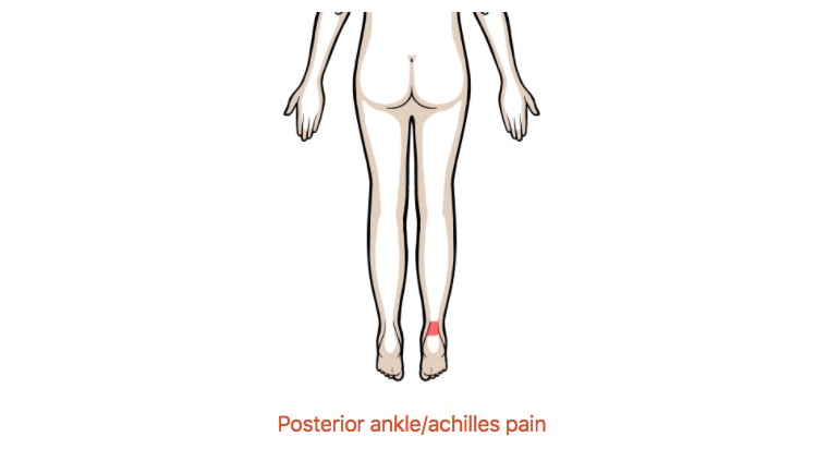
Posterior ankle/achilles pain

Patients with varying degrees of achilles tendon pathology making presentation and potential treatment differ slightly; however, they will usually present with fairly localized pain in the achilles tendon and toward the heel!

For more clinical findings click here!

Anatomy

Image via Complete Anatomy by 3D4 Medical

Image via Complete Anatomy by 3D4 Medical

Special Tests

The arc sign is and excellent assessment to help rule in presence of achilles tendon pathology due to it’s high specificity rating!(Click image to watch 1-2 minute video)

The arc sign is and excellent assessment to help rule in presence of achilles tendon pathology due to it’s high specificity rating!

(Click image to watch 1-2 minute video)

Treatment

Insufficient dorsiflexion can be a contributing factor to achilles irritation. If this motion is limited, the achilles can experience increased loading eventually leading to tissue disruption. The gastrocnemius muscle can be a source of movement res…

Insufficient dorsiflexion can be a contributing factor to achilles irritation. If this motion is limited, the achilles can experience increased loading eventually leading to tissue disruption. The gastrocnemius muscle can be a source of movement restriction and should be addressed! (Soft tissue mobilization can be executed in this position as well)

(Click image to watch 1-2 minute video)

Therapeutic Exercise

The gastrocnemius (also achilles tendon) is subject to the highest (eccentric) torque demand during gait, which is further increased while running! As other impairments are being addressed, you may want to consider improving eccentric loading capaci…

The gastrocnemius (also achilles tendon) is subject to the highest (eccentric) torque demand during gait, which is further increased while running! As other impairments are being addressed, you may want to consider improving eccentric loading capacity. (Click image to watch 1-2 minute video)

Posterior Tibialis Tendinopathy

Muscle Power Deficits

Medial ankle and foot (diffuse) pain

Patients with posterior tibialis tendinopathy typically present to clinic with diffuse medial ankle/foot pain often with insidious onset. In more severe cases pain can also radiate into the medial calf region!

For more clinical findings click here!

Anatomy

Image via Complete Anatomy by 3D4 Medical

Image via Complete Anatomy by 3D4 Medical

Midfoot Mobility

Remember, during patient assessment, it is imperative to examine up and down the kinetic chain in order to gain a more well-rounded understanding of the patient’s pathology. That being said, mid foot mobility is still something that should be addres…

Remember, during patient assessment, it is imperative to examine up and down the kinetic chain in order to gain a more well-rounded understanding of the patient’s pathology. That being said, mid foot mobility is still something that should be addressed!

(Click image to watch 1-2 minute video)
Posterior Tibialis Assessment

In order to better rule in the possibility of posterior tibialis tendinopathy, the therapist should elongate, contract, and palpate the muscle belly and tendon looking for reproduction of symptoms. Here is an example of how to properly elongate!&nbs…

In order to better rule in the possibility of posterior tibialis tendinopathy, the therapist should elongate, contract, and palpate the muscle belly and tendon looking for reproduction of symptoms. Here is an example of how to properly elongate!   (Click image to watch 1-2 minute video)

Treatment

Often, lack of dorsiflexion range of motion can result in compensatory mechanisms through the midfoot contributing to irritation of the posterior tibialis tendon. A high-velocity low amplitude thrust to the talocrural joint can help improve this ran…

Often, lack of dorsiflexion range of motion can result in compensatory mechanisms through the midfoot contributing to irritation of the posterior tibialis tendon. A high-velocity low amplitude thrust to the talocrural joint can help improve this range of motion deficit! (Click image to watch 1-2 minute video)

Therapeutic Exercise

Motor coordination both proximally and distally can contribute to excessive pronation. A squat assessment can also double as a therapeutic exercise by cueing the patient to maintain a neutral mid foot position while performing the movement! (Click i…

Motor coordination both proximally and distally can contribute to excessive pronation. A squat assessment can also double as a therapeutic exercise by cueing the patient to maintain a neutral mid foot position while performing the movement! (Click image to watch 1-2 minute video)