Anatomy Series

Medial/Lateral Meniscus Tear

 Knee Pain with Mobility Deficits

Medial Knee Pain

Medial or lateral meniscus pathologies can present with pain planting and twisting during sporting activities or locking and clicking during gait. Meniscus tears are commonly caused by either hyperflexion or hyperextension injuries. They also can present due to degenerative changes over time. Look below for some things to consider!

If you do not know the common clinical findings no problem! Click here

Anatomy

Image via Complete Anatomy 2018 by 3D4 Medical

Image via Complete Anatomy 2018 by 3D4 Medical

Special Tests

Thessalys test is a specific test to rule in meniscal pathology. It is more specific for both medial and lateral meniscus compared to McMurry’s test. When in doubt, use Thessaly’s to assess if your patient has a meniscal pathology! (Click image to w…

Thessalys test is a specific test to rule in meniscal pathology. It is more specific for both medial and lateral meniscus compared to McMurry’s test. When in doubt, use Thessaly’s to assess if your patient has a meniscal pathology! (Click image to watch 1-2 minute video)

Treatment

Restoring knee ROM is key in patients who present with this issue. It is important to gain full knee extension before you start to work on knee flexion. This is because humans need 0-5 degrees of knee extension to walk with proper gait mechanics. En…

Restoring knee ROM is key in patients who present with this issue. It is important to gain full knee extension before you start to work on knee flexion. This is because humans need 0-5 degrees of knee extension to walk with proper gait mechanics. Ensure your patient has full knee range of motion before working on strengthening and other higher level activities.

Therapeutic Exercise

It is important to choose a home exercise for your patient that matches the treatment given. In this instance, we chose to work on knee extension. The above stretch focuses on helping the patient gain full extension of the knee. Once full range of m…

It is important to choose a home exercise for your patient that matches the treatment given. In this instance, we chose to work on knee extension. The above stretch focuses on helping the patient gain full extension of the knee. Once full range of motion of the knee has been achieved it is important to work on strengthening and higher level activities. (Click image to watch 1-2 minute video)

Patellofemoral Pain Syndrome

Patellofemoral/Anterior Knee Pain

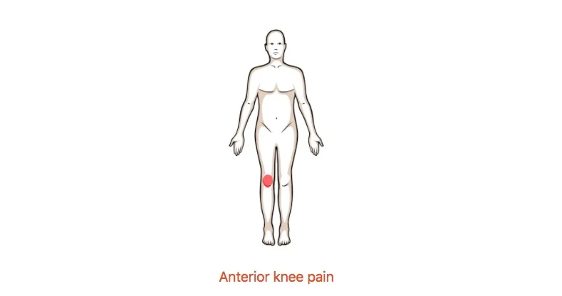
Anterior knee pain

Patella femoral pain is one of the most common problems found in physical therapy clinics. This pain pattern presents with pain behind the knee cap and pain with repetitive sporting activities such as squatting and jumping. Look below for some things to consider!

If you do not know the common clinical findings no problem! Click here

Anatomy

Image via Complete Anatomy 2018 by 3D4 Medical

Image via Complete Anatomy 2018 by 3D4 Medical

Common Movement Fault

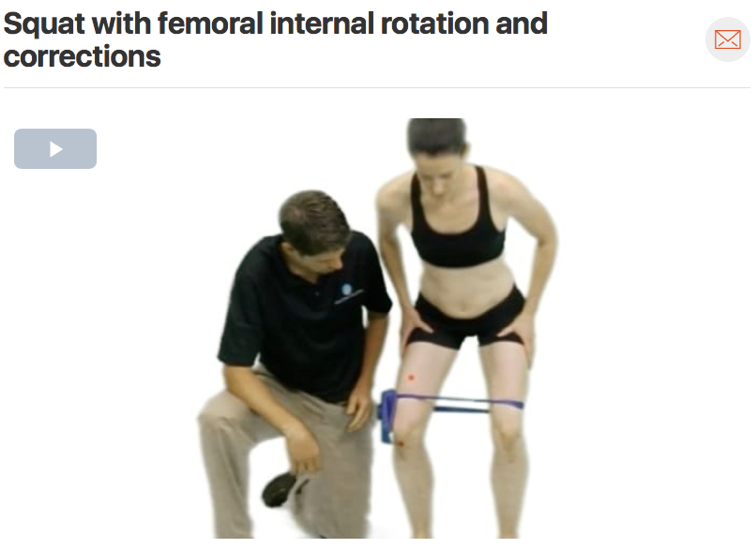
If you suspect patella femoral knee pain in your patient consider watching your patient squat and check for any movement faults. Commonly you will see your patient’s knees dive in (valgus), which we call the adduction/internal rotation movement faul…

If you suspect patella femoral knee pain in your patient consider watching your patient squat and check for any movement faults. Commonly you will see your patient’s knees dive in (valgus), which we call the adduction/internal rotation movement fault. This movement puts more stress through the knee joint which overtime can cause anterior knee pain.

Special Tests

The eccentric step down test is a provocative test to attempt to bring on the patient’s anterior knee pain. This test is great because it doubles as a movement analysis. Assess the patient for quality of movement as well as seeing if the movement br…

The eccentric step down test is a provocative test to attempt to bring on the patient’s anterior knee pain. This test is great because it doubles as a movement analysis. Assess the patient for quality of movement as well as seeing if the movement brings on the patient’s pain. (Click image to watch 1-2 minute video)

Treatment

Ensure your patient has full knee range of motion before working on strengthening and other higher level activities. A stiff rectus femoris muscle is a common impairment with patella femoral pain syndrome.

Ensure your patient has full knee range of motion before working on strengthening and other higher level activities. A stiff rectus femoris muscle is a common impairment with patella femoral pain syndrome.

Therapeutic Exercise

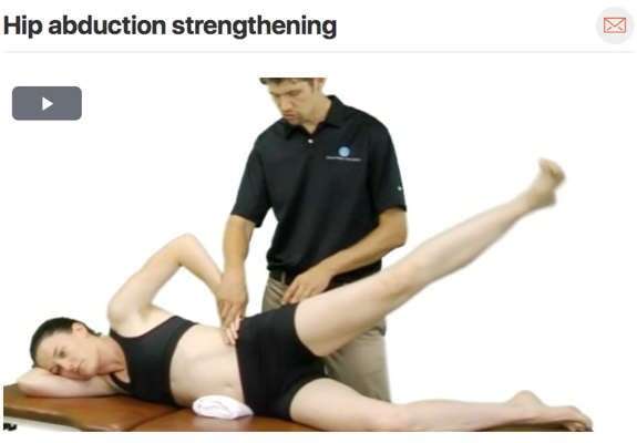
Once full range of motion of the knee has been achieved it is important to work on strengthening of both the gluteus medius and maximus muscles. These muscles control the movement of the femur and when trained can prevent the femur from adducting an…

Once full range of motion of the knee has been achieved it is important to work on strengthening of both the gluteus medius and maximus muscles. These muscles control the movement of the femur and when trained can prevent the femur from adducting and internally rotating. Both motor control and muscle strength are important to target with patella femoral pain syndrome. (Click image to watch 1-2 minute video)

Clinical Pattern Recognition: Hip Pain

From Anatomy to Discovering the Patient!

Hip pain.jpg
  • Hip pain associated with osteoarthritis (OA) is the most common cause of hip pain in older adults. Prevalence studies have shown that the rates for adult hip OA range from 0.4% to 27%.
  • Nonarthritic hip joint pain may be related to numerous underlying causes, such as femoroacetabular impingement, structural instability, acetabular labral tears, osteochondral lesions, loose bodies, ligamentum teres injury, and septic conditions
    • Labral tears in up to 20% of athletes with groin pain

    • Cam deformities more common in men (20%) than women (5%)

    • Pincer deformities more common in women (19%) than men (15%)

    • Prevalence of dysplasia in new presenters with hip pain: 32%

    • Labral tears have been observed in up to 96% of older individuals

See more prevalence information in the Clinical Pattern Recognition: Orthopaedics app

Meet the 7 common hip pain patients from the Hip Mobility Deficits Clinical Practice Guidelines!  And Non-Arthritic Hip pain Clinical Practice Guidelines + More!


Clinical Pattern Recognition

Click on the pain pattern to learn about the patients and develop your clinical patterns!

1.     Hip OsteoarthritisHip pain and mobility deficits (1 minute video | Step-by-Step Guide)

2. Adductor strainHip pain and muscle power deficits (1 minute video)


3. Hamstring strainHip pain and muscle power deficits (1 minute video | Step-By-Step Guide)


4. Hip flexor strainHip pain and muscle power deficits (1 minute video)

5. Labral tearNon-arthritic hip pain (1 minute video | Step-by-Step Guide)


6. Trochanteric bursitisLateral hip pain (1 minute video)


7. Piriformis syndrome- Hip pain with radiating leg pain (1 minute video | Step-by-Step Guide)

Piriformis Syndrome

 Hip Pain with Radiating Pain

Do you or a family member have pain in your bottom that shoots down your leg? Do you have pain down your leg after sitting for long periods of time? That pain pattern is similar to that of piriformis syndrome. Look below for some things to consider!

o   If you do not know the common clinical findings no problem! Click here


Anatomy

      Image via Complete Anatomy 2018 by 3D4 Medical

      Image via Complete Anatomy 2018 by 3D4 Medical

Common Movement Fault

A common movement pattern seen in patients that suffer from piriformis syndrome is adduction and internal rotation of the femur. This movement can occur many times through the day going down stairs, squatting, sitting cross legged, or even sleeping …

A common movement pattern seen in patients that suffer from piriformis syndrome is adduction and internal rotation of the femur. This movement can occur many times through the day going down stairs, squatting, sitting cross legged, or even sleeping on your side and allowing your knee to fall to the bed without putting a pillow between your legs. It is always important to watch how people move! (Click image to watch 1-2 minute video)

Special Tests

A good clinical test to consider when evaluating someone you suspect has piriformis syndrome is the FAIR test. Which is used to rule in/rule out piriformis syndrome by putting the piriformis on stretch. The straight leg raises test can also be useful to determine if there is any neural involvement contributing to the lower extremity pain. (Click image to watch 1-2 minute video. LEFT | RIGHT)

Treatment

Piriformis syndrome can be caused by the sciatic nerve getting entrapped in the piriformis muscle. It is important to work on the entrapment site and then follow up with nerve glides to increase neural mobility. (Click image to watch 1-2 minute video. LEFT | RIGHT)

Therapeutic Exercise

Therapeutic exercises chosen should always closely match treatment given in clinic. It is also important to correct the movement fault to prevent future irritation of the tissue source! This video shows the therapist correcting the patients squattin…

Therapeutic exercises chosen should always closely match treatment given in clinic. It is also important to correct the movement fault to prevent future irritation of the tissue source! This video shows the therapist correcting the patients squatting form. (Click image to watch 1-2 minute video)

Acetabular Labrum Tear

Hip Pain with Movement Coordination Impairments

Screen Shot 2018-03-14 at 7.41.40 AM.jpg

Occasionally mistaken for an adductor muscle strain, patients with hip labral tears will present with disabling anterior hip or groin pain often associated with a clicking sound or sensation. For more clinical findings click here!

Anatomy

      Image via Complete Anatomy 2018 by 3D4 Medical

      Image via Complete Anatomy 2018 by 3D4 Medical

Common Movement Fault

If you suspect labral pathology consider assessing your patient for an anterior humeral movement fault. If the head of the humerus more readily glides anteriorly this can cause excessive stress to the anterior portion of the labrum. Prone hip extens…

If you suspect labral pathology consider assessing your patient for an anterior humeral movement fault. If the head of the humerus more readily glides anteriorly this can cause excessive stress to the anterior portion of the labrum. Prone hip extension is one way the therapist can assess for anterior humeral glide. (Click image to watch 1-2 minute video)
Special Tests

The hip scour test can be useful during assessment to help rule out potential labral tear or degeneration! (Click image to watch 1-2 minute video)

The hip scour test can be useful during assessment to help rule out potential labral tear or degeneration! (Click image to watch 1-2 minute video)
Treatment

Restriction of hip posterior glide mobility can also contribute to imprecise movement of the head of the femur in the acetabulum potentially exacerbating the anterior femoral glide movement fault. Hip flexion mobilization with movement can help redu…

Restriction of hip posterior glide mobility can also contribute to imprecise movement of the head of the femur in the acetabulum potentially exacerbating the anterior femoral glide movement fault. Hip flexion mobilization with movement can help reduce this posterior glide restriction.  (Click the image to watch 1-2 minute video)

Therapeutic Exercise

A good therapeutic exercise to follow hip flexion mobilization with movement would be the quadruped rock back; however, after mobility is improved, it is important to address gluteal strength along with motor control of the lower extremity! (Click image to watch 1-2 minute videos left | right)